
중요한 뇌혈관질환에는 어떤것이 있을까?
- STROKE(뇌졸중, aka 중풍) / Cerebral infarction
- Subarachnoid hemorrhage(SAH, 지주막하출혈)
> Trauma
> Aneurysm
> AVM
- Intracranial hemorrhage(ICH)
.
.
- moyamoya disease
- CNS vasculitis
- Dissection
리스트에 있는 혈관질환들의 이름들을 보면 상당히 무시무시한 이름들이 많은 것을 알 수 있다.
이런 혈관질환들을 평가, 진단하려면 어떻게 해야할까? CT angiography, MR angiography, digital subtraction angiography와 같은 혈관조영술 영상으로 뇌혈관의 이상을 평가할수 있겠다. 혈관조영술은 조영제를 사용해 혈관을 평가한다.
CT angiography나 MR angiography의 indication은 다음과 같은것들이 알려져 있다.
- Acute SAH
- Cerebral aneurysm or AVM without acute SAH
- TIA/stroke
- brain tumor: blood supply 평가
- vasculitis
- cerebral artery stenosis 평가
- cerebral venous thrombosis
- 각종 vascular anomalies(moyamoya)
혈관조영술로 혈관을 평가하기 위해선 뇌혈관에 대해 알고 있어야한다.
뇌혈관 영상해부학에 대해 알아보자
Vascular anatomy
- Anterior circulation
- Internal carotid artery
- Anterior cerebral artery
- Middle cerebral artery
- Circle of Willis
- Posterior circulation
- Vertebral artery
- Branches of vertebro-basilar system
- Basilar perforators to brainstem
- Posterior cerebral artery
- Thalamoperforators

- Circle of willis: ACA, Acom, PCA, Pcom, ICA
-> 교수님들이 맨날 물어보는 왕족보, 국룰 , 시험에도 많이 나올것이다.
-> 85%의 saccular aneurysm이 circle of willis에서 발생한다고 흔히 알려져 있다. Anterior communicating artery(35%), internal carotied artery(30%), Middle cerebral artery, basilar artery bifurcation이 aneurysm의 호발 부위다.
(Keedy A. An overview of intracranial aneurysms. Mcgill J Med. 2006;9(2):141-146.)
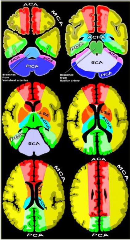
Vascular territory
- Anterior cerebral artery(ACA): medial part of frontal and parietal lobe, anterior portion of corpus callosum, basal ganglia, internal capsule
- Middle cerebral artery(MCA): lateral surface of hemisphere, except for the medial part of the frontal and the parietal lobe(ACA cover)/ inferior part of temporal lobe(PCA),
- Posterior cerebral artery(PCA):
- Posterior thalamoperforating arteries: midbrain/thalamus
- Cortical branch: infereromedial part of the temporal lobe, occipital pole, visual cortex, splenium of the corpus callosum
- Superior cerebellar artery(SCA): superior and tentorial surface of the cerebellum
- Anterior inferior cerebellar artery(AICA): lateral side of the cerebellum
- Posterior inferior cerebellar artery(PICA): inferior occipital surface of the cerebellum
- Verebral / basilar artery: medulla oblongata and the pons
Vascular territory를 이해하는 것은 stroke/TIA를 평가하는데 중요하다.
KMLE와 학교시험에도 자주 나오는 부분이다. 임상증상을 통해 어떤 뇌 부위가 손상받았는지 맞추는 문제가 주로 나온다.
- ACA infarction:
- contralateral sensory and motor symptom in the lower extremity, sparing hands and face
- incontinence (bilateral lesion)
- Left sided lesion: (typically) akinetic mutism / transcortical motor aphasia
- right sided infarction: (typically) confusion / motor hemineglect
ACA infarction의 경우 하체에 더 심한 운동장애가 나타나는 것이 특징이다.
- MCA infarction: stroke가 가장 많이 호발하는 혈관이다.
- (typically) hemiparesis, facial plegia, contralateral sensory loss
- Neurological deficitis affecting the face and upper extremity more than the lower extremity
- dominant hemisphere involvement: aphasia(receptive, expressive, or both)
- nondominant hemisphere: inattention, neglect, extinction on double-simultaneous stimulation, dysarthria without aphasia, constructional apraxia
- homonymous hemianopsia and gaze preference toward the side of the infarct
MCA infarctinon 은 상체에 더 심한 운동장애, 실어증, gerstmann syndrome, 편측 무시, 반맹이 국룰 처럼 알려지고 있다.
- PCA infarction
- ataxia, nystagmus, vertigo
- unilateral limb weakness, dizziness, gait ataxia, headache, dysarthria
- visual field loss, blurry vision: contralateral homonymous hemianopsia
- Veretebral artery
- Medulla 침범 증상: ipsilateral horner's syndrome, cranial nerve V symptom, unilateral limb ataxia...
- Cerebellar infarction
- dizziness(with or without vertigo), nausea, vomiting, gait instability, headache, limb ataxia
- dysmetria
- nystagmus
- intractable hiccups
- cerebellar edema -> obstructing hydrocephalus or symptomatic mass effect(응급)
- Lacunar infarction
- pure motor on sensory deficits caused by infarction of small penetrating arteries
Venous anatomy
- superficial venous system
- Superior cerebral vein -> Superior sagittal sinus
- Superficial middle cerebral vein
- Sup. anastomosing vein of Trolard -> superior sagittal sinus
- Inf. anastomosing vein of Labbe -> transverse sinus
- Inferior cerebral vein
- deep venous system
- internal cerebral vein -> vein of galen
- Deep middle cerebral vein -> basal vein of rosenthal -> vein of glaen
- dural sinus
cortical venous drainage와 deep venous drainage로 나뉘고 합쳐져서 dural sinus로 배출됨

'DOC > 영상의학' 카테고리의 다른 글
| 뇌 영상해부학 (brain anatomy), MRI 보는법 (0) | 2022.03.04 |
|---|---|
| Chest x-ray 보는법 (3): lobal anatomy and atelectasis (0) | 2022.03.04 |
| Chest x ray 보는법 (2): anatomy and search pattern (0) | 2022.03.04 |
| Chest x ray 보는법 (1): chest AP vs PA (0) | 2022.03.04 |